Torn Retinaculum Ankle : Peroneal Tendon Subluxation: Treatment, Recovery ... : He came back after 2 weeks and reported that he had a left ankle injury when playing basketball and was using crutches and was on air boots when came to see me.
Torn Retinaculum Ankle : Peroneal Tendon Subluxation: Treatment, Recovery ... : He came back after 2 weeks and reported that he had a left ankle injury when playing basketball and was using crutches and was on air boots when came to see me.. Up to date literature mostly describes proximal peroneal tendon dislocations due to superior peroneal retinaculum (spr) tear. Occasionally, the covering that holds the peroneus tendons behind the lateral malleolus (the retinaculum) can be loose or torn and the tendons can snap back and forth out of their normal grooves, this snapping sensation is felt by the patient and can causes further stress/friction on the tendons. Moderate to severe pain in the ankle and foot with ambulating or running or in fact any movement of the foot. The main symptoms of inferior extensor retinaculum pain are: The symptoms of subluxation may include:
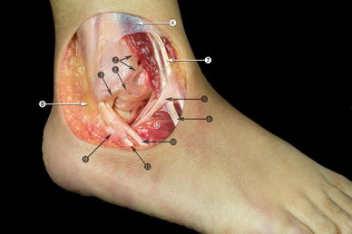
A retinaculum is a band of thick deep fascia that holds the long tendons of your ankle (those that cross the ankle) in place. Moderate to severe pain in the ankle and foot with ambulating or running or in fact any movement of the foot. Plastic surgery 52 years experience. Once the cast is off, a strengthening program is prescribed by a physiotherapist to help you return back to normal function. Patient had an ankle injury and the retinaculum over the peroneal brevis and longus was torn.
Anatomy of the ankle ligaments: a pictorial essay ... from media.springernature.com Any damaged tendon is removed prior to repair. Dr yuranga weerakkody and dr henry knipe et al. Again a previously torn retinaculum will be thickened and ill defined compared to a normal retinaculum which is usually thin and well defined. The stem, or frondiform, ligament; The forceful stretch on the peroneals can rip the retinaculum that keeps the peroneal tendons positioned in the groove. Usually, this is when the foot is forced up and to the side (for example, a caught ski tip.) causes of peroneal tendon injuries During the typical inversion ankle sprain, the foot rolls in. Ankle ext retinaculum torn after fasciotomy & injury in pt.
The physician repaired the retinaculum which holds these down.
Any damaged tendon is removed prior to repair. Retinaculum are a major source of neurological receptors involved in balance and proprioception. And the oblique superolateral band 6 . He came back after 2 weeks and reported that he had a left ankle injury when playing basketball and was using crutches and was on air boots when came to see me. Up to date literature mostly describes proximal peroneal tendon dislocations due to superior peroneal retinaculum (spr) tear. The forceful stretch on the peroneals can rip the retinaculum that keeps the peroneal tendons positioned in the groove. Depending on the severity of tear, sometimes surgery is needed to repair the retinaculum. In other cases, subluxation occurs following trauma, such as an ankle sprain. A mass over the anterior aspect of the ankle most likely represents the retracted proximal tendon stump as it becomes entrapped at the extensor retinaculum (24). Your doctor may also order a magnetic resonance imaging (mri) scan of your ankle. Damage or injury to the tissues that stabilize the tendons (retinaculum) can lead to chronic tendon subluxation. Retinaculum also acts as a pulley system increasing mechanical advantage. This is called an avulsion fracture.
This is called an avulsion fracture. Damage or injury to the tissues that stabilize the tendons (retinaculum) can lead to chronic tendon subluxation. The main symptoms of inferior extensor retinaculum pain are: Retinaculum also acts as a pulley system increasing mechanical advantage. The forceful stretch on the peroneals can rip the retinaculum that keeps the peroneal tendons positioned in the groove.
Stock Trial Exhibits from cdn.shopify.com Previously torn extensor retinaculum of ankle which is now markedly thickened and irregular (blue arrows). The tendon sheaths and retinaculum (structure that binds tendons down) are repaired. And the oblique superolateral band 6 . During the typical inversion ankle sprain, the foot rolls in. The forceful stretch on the peroneals can rip the retinaculum that keeps the peroneal tendons positioned in the groove. Some of the symptoms of strained flexor retinaculum of the foot are: Up to date literature mostly describes proximal peroneal tendon dislocations due to superior peroneal retinaculum (spr) tear. Any damaged tendon is removed prior to repair.
During the typical inversion ankle sprain, the foot rolls in.
Official fit assistance for canyon speedmax cfr, cf slx, and cf (disc & rim brake) Medial flexor retinaculum injury is often also associated with superficial deltoid pathology and/or medial malleolar fracture. This is called an avulsion fracture. Damage or injury to the tissues that stabilize the tendons (retinaculum) can lead to chronic tendon subluxation. And the oblique superolateral band 6 . It is a complex structure that has four components: Damage or injury to the tissues that stabilize the tendons (retinaculum) can lead to chronic tendon subluxation. A sprain that injures the ligaments on the outer edge of the ankle can also damage the peroneal tendons. How can reconstruct retinaculum w/o causing cs pressure to return? answered by dr. The symptoms of subluxation may include: Usually, this is when the foot is forced up and to the side (for example, a caught ski tip.) causes of peroneal tendon injuries 1 doctor answer • 1 doctor weighed in. He said that the temporary insoles were ok to ease the pain in the bottom of the foot but achille's tendon still hurting and pain on the lateral border hasn't changed.
A sprain that injures the ligaments on the outer edge of the ankle can also damage the peroneal tendons. Tendon tear is repaired with suture and tendon is returned to tubular shape. Again a previously torn retinaculum will be thickened and ill defined compared to a normal retinaculum which is usually thin and well defined. Up to date literature mostly describes proximal peroneal tendon dislocations due to superior peroneal retinaculum (spr) tear. Some of the symptoms of strained flexor retinaculum of the foot are:
Radiology Cases: Transient Patella Disclocation from 1.bp.blogspot.com However, the physician wants to bill for repair of dislocating tendon (27675) but in addition wants to bill for repairing the tendons themselves (27659) which i don't understand. The symptoms of subluxation may include: And the oblique superolateral band 6 . Retinaculum are a major source of neurological receptors involved in balance and proprioception. This is called an avulsion fracture. How can reconstruct retinaculum w/o causing cs pressure to return? The stem, or frondiform, ligament; A snapping feeling of the tendon around the ankle bone
Some of the symptoms of strained flexor retinaculum of the foot are:
Plantaris tendon (if you have one) overlay may do the. The main symptoms of inferior extensor retinaculum pain are: Bmc (time machine) official bmc time machine owners thread; The symptoms of subluxation may include: The physician repaired the retinaculum which holds these down. During the typical inversion ankle sprain, the foot rolls in. The tears are typically vertical, and a complete rupture is rare. Tibialis anterior tendon (arrowheads) is most medially located and passes though tunnels formed by oblique superomedial limb (short arrows) and oblique inferomedial limb (long arrows) of. Tendon tear is repaired with suture and tendon is returned to tubular shape. How can reconstruct retinaculum w/o causing cs pressure to return? answered by dr. Skin is closed with suture, followed by application of bandages and below the knee cast. As a result, the tendons can jump out of the groove. This is called an avulsion fracture.
Other causes, such as an organized hematoma or fibrous tissue surrounding the torn tendon stump or fibrosis of the synovial sheath and retinaculum, have been suggested (16) torn retina. The physician repaired the retinaculum which holds these down.


0 Comments